Services
Ultrasound
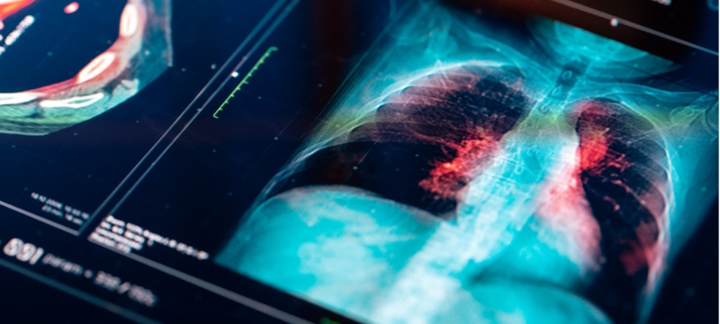
An ultrasound, or sonography, uses high-frequency sound waves to create images and capture movements of internal organs, tissues, structures, and blood flow in real time. The sound waves create a visual image that can be used to assess health and disease, gauge the effectiveness of medical treatment, and provide image guidance for needle biopsies.
What types of ultrasounds do we offer?
Ultrasound imaging is a noninvasive test that uses a transducer wand rolled over gel on the skin to create 3D quality images on a computer screen. Ultrasound captures internal images and movements in real-time, blood flow, and abnormalities in organs and structures.
A breast ultrasound can provide information about lumps and abnormalities discovered during a screening or diagnostic mammogram, physical exam or other test.
Cardiac ultrasound can spot places in the heart muscle that are damaged or working ineffectively, locate injury from a previously diagnosed or undiagnosed heart attack, and isolate details about blood flow, possible clots, excess fluid around the heart and more.
A fine needle aspiration biopsy of the Thyroid is a procedure that removes a small sample of tissue from your thyroid gland. Cells are removed through a small, hollow needle.
Know what to expect from your visit
During your exam
- Lie still on your back on the examination table.
- A clear gel will be applied to the examination area.
- Your technologist or radiologist will sweep the transducer wand firmly back and forth on the examination area.
- The applied pressure may cause mild discomfort.
- The examination usually takes less than 30 minutes.
ACR Accredited in Breast Ultrasound
Our facilities have been accredited by the American College of Radiology (ACR) for accuracy, safety and best practice standards in Breast Ultrasound.